The proteins that fix (almost) everything
Proteins can make any inventor green with envy. It is proteins that make the body work. But when these same super-substances make mistakes, we may get sick with things like cancer or Alzheimer's disease. The job of researchers is to sort out the proteins when they malfunction.
Text: Annika Lund for the magazine Medicinsk Vetenskap no 4 2022 / Spotlight on proteins
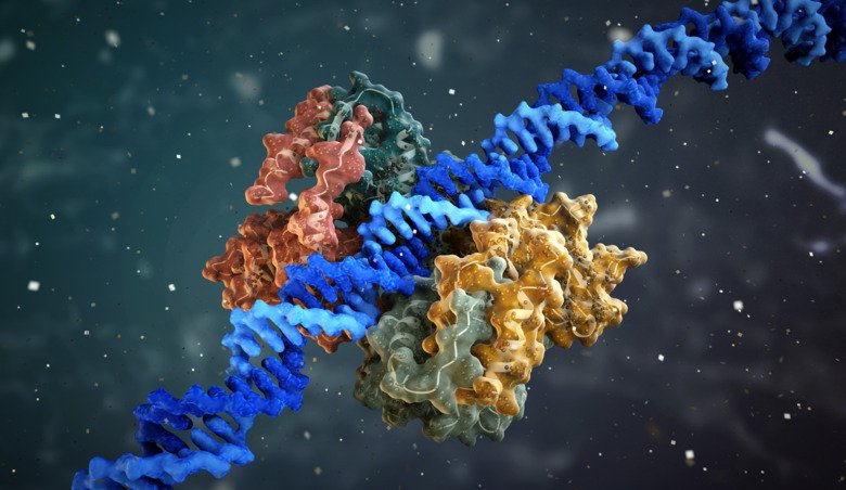
If you google the phrase “building blocks of the body” you will quickly realise it is an overused and vague expression. More often than not, the metaphor is applied to the molecules that help the body's cells function: proteins. But they are so much more than just building blocks.
So – what exactly is a protein? Is there a definition?
Well, all proteins are made up of amino acids that join together to form a chain that folds into a particular structure. Otherwise, they are different – very different. Because if you ask what proteins do in the body, the answer is a little all over the place (Check out the protein school at the bottom for a more precise answer).
The great leaps of protein research
Protein research has made almost unimaginable strides in the last two decades. A first baby step was taken when researchers succeeded in mapping the human genome. They found that on the three-metre-long strand of DNA found in every cell, there are around 21,000 sections that produce proteins, the protein-producing genes.

The researchers then realised that each gene can produce several versions of each protein. That makes about 90,000 protein variants. And the proteins can change even after they are made, for example by another molecule attaching to them. This affects the proteins in several ways, explains Janne Lehtiö, Professor of Proteomics at the Department of Oncology-Pathology at Karolinska Institutet.
“The chemical modifications that occur in proteins after they have been formed affect where they will be in the cell, how stable they are and which other proteins they interact with. All this affects their function.”
He describes it all with a picture, in which a tree that makes plums is likened to a gene that makes a protein.
“We can imagine that the plums come from the same tree, but still, the plums in a cake are not the same as a dried plum in a pork roast. Similarly, a gene can produce protein variants that have different accessories and thereby different functions. Right now, we know relatively little about these changes that occur after the protein is created, referred to as post-translational modifications. We do not really know much about how they work in biological systems,” says Janne Lehtiö.
Overall, it is difficult to say how many proteins there actually are in the body. So the 21,000 or so genes make about 90,000 protein variants – but if you add all the variants that arise as a result of later changes, the number is not known.
Proteome researchers study the whole protein picture
That is exactly what proteomic researchers are trying to study – the whole protein picture. Per cell type. Or per tissue type.
“I started out as a protein chemist, looking at individual proteins, how their structure changes and how that controls their function. But I eventually understood that proteins are influenced by their environment and work in teams. The biology can only be understood by looking at all the proteins at the same time, and that is what we do in proteomics. You could say that a protein chemist focuses on a violinist, but a proteomic scientist tries to listen to the whole symphony orchestra,” says Janne Lehtiö.
Among other things, he and his colleagues are trying to create reference proteomes for different cell types, i.e. which proteins should “normally” be included in a healthy cell of a certain type. This reference proteome can then be compared with what is present in diseased cells of the same type. For example, researchers have tried to get a picture of what proteins are usually present in healthy cells of the type that can develop into lung cancer. They can then compare this with the protein picture in cancer cells and gain a better understanding of how the disease develops and what drives it.
Janne Lehtiö's research group is also looking at the protein pattern in HER2-positive breast cancer, which occurs when multiple copies of the gene that makes the HER2 protein are made. This causes the cell to produce a lot of HER2, which tells the cell to divide. A large amount of this protein leads to massive cell division and a tumour develops.
There are drugs that can lock onto the HER2 protein, which can then no longer send signals to divide. These drugs have made a big difference to breast cancer care, curing many patients.
May affect cancer treatment choices
But in some patients, the cancer cells survive anyway. After a few good whacks from the drug, the tumours grow back and the disease is rediscovered.
Janne Lehtiö and his colleagues have devoted themselves to trying to understand what separates patients who suffer relapse from those who are cured. They have been working with a team of doctors on a study involving 150 patients with HER2-positive breast cancer. The researchers mapped the proteome in the cancer cells of all these patients, both before starting treatment and after two weeks.
This has provided interesting answers.
“You could say that blocking the HER2 protein in these patients silences a violinist who is playing out of tune. But if you listen to the whole orchestra, you may find that more are playing out of tune. Some of these patients have more proteins that do not cooperate and should be stopped,” says Janne Lehtiö.
The researchers have found abnormalities that are recognisable from other cancers and are treated with completely different drugs, which are not currently used in HER2-positive breast cancer.
“We are currently optimising our method in order to be able to select patients who have certain changes that we believe are treatable. At the same time, we are discussing how this could lead to a clinical trial, where a subgroup of patients with HER2-positive breast cancer could receive a cocktail of several already approved drugs,” says Janne Lehtiö.
Many cancer cells lack p53
There are some proteins that are relevant to a great number of cancers, regardless of the organ in which the tumours originate. One of these is p53 – the most studied protein in the world. It is normally found in all cells and is often likened to a superhero, the Guardian of the Genome, constantly monitoring the cell's DNA. If DNA damage is detected, p53 acts by preventing the cell from dividing. This eventually leads to the death of the cell as well.
So a cell lacking p53 has lost an important emergency brake. Many cancer cells have no or too little p53.

“Cancer cells have a range of defects, so changes involving p53 are rarely the only problem. But it is a defect that makes all other defects irrelevant. Up to 60 percent of all tumours have mutations that affect p53,” says Michael Landreh, Associate Professor at the Department of Microbiology, Tumour and Cell Biology at Karolinska Institutet.
He has spent a lot of time looking at p53 and MYC, another protein relevant to many cancers. MYC can be said to “hijack” a cell and make it produce proteins ruthlessly in order to protect only itself - the hijacked cell becomes a cancer cell.
A shared trait of p53 and MYC proteins is that they are unstructured – they have no easily described shape. MYC has been called undruggable, unreachable for treatment, for this reason – it is hard to find a place on the protein where you can get something to adhere, something that might change it so that it stops behaving so harmfully.
“In the case of p53, part of the protein has a distinct structure that binds to the DNA molecule. But the rest of the protein consists of a large number of wispy strands that flutter around like cooked spaghetti, in constant motion,” explains Michael Landreh.
So whoever wants to interact with p53 has to somehow get things to adhere to that cooked spaghetti. But what adheres there? And how can you tell if something has adhered?
Michael Landreh is investigating this with mass spectrometry, a method that can describe the mass of a protein with great precision. In simple terms, the researchers shoot the protein into a tunnel and see how fast it flies, which is related to its weight. Then they expose the protein to different molecules, such as other proteins or drugs, and see if it flies slower in the tunnel. If it does, it has become heavier – something has bound to it.
“We want to understand how these proteins and lots of other proteins interact with each other. We want to know which ones bind to each other or vice versa – what might make one protein let go of another. The goal is to be able to influence harmful proteins and make them harmless,” says Michael Landreh.
Malfunctioning protein in Alzheimer's disease
But diseases can also occur because a protein interacts with itself in a harmful way. This is the case, for example, in Alzheimer's disease, where a very small protein, called a peptide, malfunctions. It is beta-amyloid, which is also found in healthy brains, that folds in the wrong way. You could say that the peptide folds up like a folding rule again and again, until it has formed a so-called fibril, a strong cable of proteins or peptides. It is a tough structure that is very difficult to dissolve. They can also clump together and form plaques.
Fibrils and plaques can be formed from different kinds of proteins and peptides. They are present in the brain in several neurodegenerative diseases, such as Alzheimer's disease and Parkinson's disease.
However, the role of these fibrils and plaques is a matter of debate.

“It seems that the very formation of the fibrils involves something toxic and harmful to the neurons. The final fibrils may mostly be some form of final storage. The major damage seems to happen during the process on the way there,” says Axel Abelein, a biophysicist and researcher at the Department of Biosciences and Nutrition at Karolinska Institutet.
He is researching the state between the normal peptide and the final fibril. In these phases, the accumulations are called oligomers (consisting of a few proteins) or protofibrils (consisting of more proteins and having started to look like fibrils). For some reason, more and more peptides are attracted to these structures, which at the same time fold incorrectly.
Axel Abelein and his colleagues are trying to prevent the formation of these toxic oligomers and protofibrils. To do this, they are investigating the function of additional proteins in this context, known as chaperone proteins, which help with the folding process.
The researchers have shown in test tubes that the process of fibril formation is slower when a certain chaperone protein, called BRICHOS, is present. They have also tried giving this chaperone protein in injection form to mice that have been bred to have Alzheimer's disease. Those experiments showed that mice that received the treatment retained more cognitive functions, such as their working memory, than mice that did not receive the treatment.
“Our goal is to better understand neurodegenerative diseases by pinpointing why certain proteins, like beta-amyloid, behave so strangely. When it comes to Alzheimer's disease, we hope to lay the foundations for a new drug, and this chaperone protein is our candidate,” says Axel Abelein.
Amino acids in a unique form
What is a protein? Basically a chain of amino acids, but it is more complicated than that
Proteins are molecules made up of chains of 20 different amino acids.
The amino acids have a specific order determined by the order of nitrogen bases in a gene.
Each protein has a unique three-dimensional structure that determines its function.
The three-dimensional structure can be described in four levels, of which the primary structure – the order of amino acids – is the simplest.
What do proteins do?
The short answer is almost everything. The long answer is that they ...
... give us structure
Proteins are components of the body's tissues, such as muscles, tendons and bones.
... provide transport
Proteins make sure the right thing goes to the right place. One example is haemoglobin, which moves oxygen around the body.
... make things happen
Enzymes, which can be said to control the speed of bodily processes, such as how quickly the food we eat is converted into things our cells can use, are also proteins.
... move
Both the contractions of muscles and the movement of cells in the body are controlled by proteins that generate movement, such as actin and myosin.
... defend us
Proteins are also important in the immune system – antibodies are proteins.
... act as stores
Proteins can store other molecules until they need to be used. One example is ferritin, which stores iron inside the body's cells.
... communicate
Many of our hormones, the body's messengers that regulate everything from growth to blood pressure and blood sugar, are proteins. They are also the receptors that receive various signals, such as the receptors in our sensory cells that allow us to experience the world around us.
A significant part of you
How many proteins are there? It is hard to know exactly, but here are some key numbers:
About 15 percent of body weight is made up of proteins.
An estimated 90,000 different kinds of proteins are produced in the body. More protein variants are then added by changing existing proteins.
It is estimated that the average cell contains tens of millions of protein molecules. The number ranges from a few hundred to thousands, depending on the protein.
Red blood cells contain a few thousand different types of proteins, while nerve cells, for example, may need a much larger number of protein types to function.
Sources: Janne Lehtiö, www.thoughtco.com, NE