Protein movement reveals brain tumour's weak spot
Proteins do not have a single shape, but are more like dynamic robots. But – how do they actually move? And what does this tell us about their function? Laura Orellana describes the movement of proteins using computer simulations – and has discovered a new drug target for glioblastoma brain tumours.

Text: Annika Lund for Medicinsk Vetenskap no 4 2022 / Spotlight on proteins
Decades of research data are collected in the Protein Data Bank, which describes the three-dimensional structure of different kinds of biological molecules, such as proteins. The knowledge is based on experimental methods such as X-ray crystallography or nuclear magnetic resonance (NMR).
“This database currently contains about 60,000 known structures from human proteins,” says Laura Orellana, a biophysicist and researcher at the Department of Oncology-Pathology at Karolinska Institutet.
But these are still images that do not show how the protein moves.
“This is a problem, because it is in the movement itself that the function lies. Proteins are like tiny nanomachines that perform all the cellular functions that keep us alive, but a still image of a dynamic machine gives a limited picture of what it does when it is working,” says Laura Orellana.
She is researching computer simulations that describe the movements of proteins. The simulations are based on existing "images" of different proteins, animated using the laws of physics.
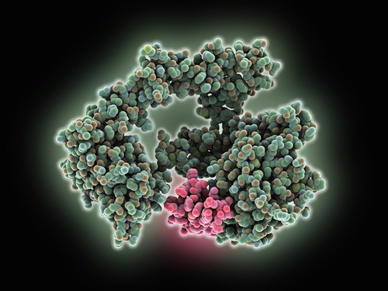
One of the proteins she is trying to simulate is the oncogene EGFR (HER1), which encourages cell division. It is a complex and dynamic protein with one part outside and one part inside the cell. In lung cancer, the mutated parts of the protein are inside the cell – but in glioblastoma, a form of brain cancer, they are outside. This in turn affects the efficacy of drugs.
Laura Orellana and her colleagues have investigated how EGFR moves in glioblastoma.
“It is like putting together a big puzzle. We want to create a true picture of how different variants of the protein as a whole move,” says Laura Orellana.
The researchers seem to have put the puzzle together well. Through simulations, they have revealed that in glioblastoma, mutations lead to the movement of a large part of the extracellular part of the protein. This activates the intracellular part in a specific way that is different from lung cancer and drives tumour development in glioblastoma. But the computer simulations have also revealed an exposed contact surface where an antibody can attach. The researchers have confirmed what they have seen in computer simulations in experiments with cell and animal models. A specific antibody has been able to bind to this particular contact surface and it has dramatically inhibited the growth of glioblastoma in cell and animal studies.
“We need to understand how this machinery moves and works at the atomic level. Drugs do not bind anywhere in proteins, but instead bind only in special pockets. Such a pocket can be open sometimes and closed sometimes, depending on the movement of the protein,” says Laura Orellana.