Scar formation after spinal cord injury is more complex than previously thought
New research reveals that scar formation after spinal cord injuries is more complex than previously thought. Scientists have identified two types of perivascular cells as key contributors to scar tissue, which hinders nerve regeneration and functional recovery. These findings, published in Natural Neuroscience, are also relevant for other brain and spinal cord injuries and could lead to targeted therapies for reducing scarring and improving outcomes.
The central nervous system (CNS) has very limited healing abilities. Injuries from traffic accidents, sports incidents, or autoimmune diseases like multiple sclerosis often lead to permanent functional deficits.
Regardless of the injury's cause, the body responds by forming a boundary around the damaged tissue, which eventually becomes permanent scar tissue.
Two contributing cell types
While scar tissue seals the damaged area, it also prevents functional repair. After spinal cord injuries, scar tissue blocks the regeneration of nerve fibers that connect the brain with the body, resulting in paralysis after severe injuries.
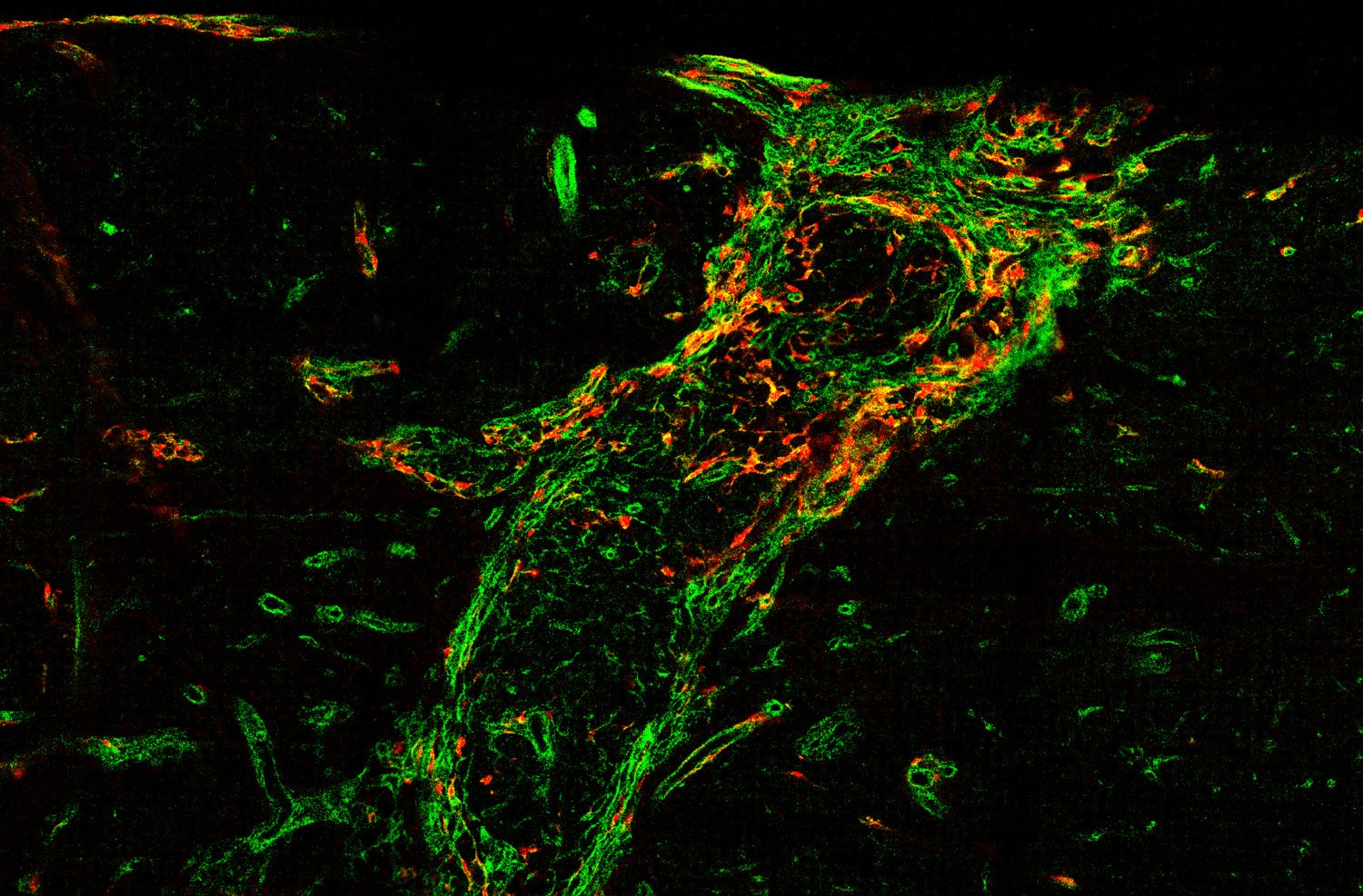
The research team led by Christian Göritz at Karolinska Institutet has made significant progress in understanding how scar tissue forms in the CNS. The group now identified two distinct types of perivascular cells, which line different parts of blood vessels, as the major contributors to fibrotic scar tissue after spinal cord injury. Depending on the lesion's location, the two identified cell types contribute differently.
“We found that damage to the spinal cord activates perivascular cells close to the damaged area and induces the generation of myofibroblasts, which consequently form persistent scar tissue,” explains first author Daniel Holl, researcher at the Department of Cell and Molecular Biology.
By examining the process of scar formation in detail, the researchers hope to identify specific therapeutic targets to control fibrotic scarring.
“Understanding the cellular origin and composition of scar tissue is a critical step in our endeavor to develop a therapy that reduces scarring and promotes functional recovery after spinal cord injury. Now we know the cells that we need to target,” notes Christian Göritz, corresponding author at the same department and head of the research team.
The findings are also relevant for other injuries to the brain and spinal cord, which lead to scar formation.
The project was suported by the Swedish Research Council, the Swedish Brain Foundation, Knut and Alice Wallenberg Foundation and Wings for Life. There are no reported conflicts of interest.
Publication
"Distinct origin and region-dependent contribution of stromal fibroblasts to fibrosis following traumatic injury in mice", Holl D, Hau WF, Julien A, Banitalebi S, Kalkitsas J, Savant S, Llorens-Bobadilla E, Herault Y, Pavlovic G, Amiry-Moghaddam M, Dias DO & Göritz C. Natural Neuroscience, online June 7 2024.