Washing proteins with soap reveals new clues for drug development
Using a trick involving detergent and mass spectrometry, a research group has been able to wash and weigh protein molecules to determine which lipids make the protein work. The findings may help design molecules that stick to individual membrane proteins and pave the way for the development of new drugs including antibiotics and cancer therapies. The study is published in Angewandte Chemie International Edition.
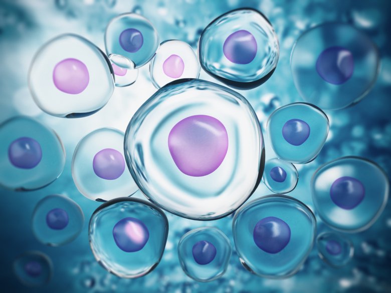
All cells are surrounded by the lipid membrane, a thin layer of fat-like molecules that separates the inside of the cell from the outside. Proteins in the membrane are surrounded by thousands of lipid molecules, but some need special lipids to work. Finding these lipids can be like finding a needle in a haystack, because every protein in the membrane makes thousands of contacts with different lipids every second.
In this study, a team of researchers from Karolinska Institutet, Oxford University, Stockholm University and Warwick University were able to show how a membrane protein can find and apprehend specific lipids. The researchers used a trick: They surrounded the protein with lipids and then added detergent, soap molecules that compete with the lipids to bind to the protein. The researchers then used a technique called native mass spectrometry to measure the weight of the proteins before and after washing, to find out which lipids remained.
Most lipids were washed away
”Our computational models show that most lipids will be washed away by the detergent, while those that make the protein work tend to stick,” says Michael Landreh, assistant professor at the Department of Microbiology, Tumor and Cell Biology at Karolinska Institutet who led the study.
The findings provide new insights into how proteins are turned on and off, says Landreh.
“We could influence proteins related to various diseases by adding or removing lipids, but in order to do so we would need to identify the right lipids,” says Landreh. “Our study shows just how the membrane protein is connected to various lipids.”
The researchers investigated MurJ, a protein bacterium use to build the cell wall.
Promising strategy for the development of new drugs
“It was exciting to see that two lipids could not be washed away from the protein: one turned out to be the raw material for the cell wall, and the other is a lipid that stops the protein from working,” says Landreh. “Additional knowledge about the connection between different lipids could pave the way for new antibiotics.”

The researchers also used the method to investigate other proteins. Presenilin, which produces molecules related to Alzheimer’s disease, did not show any lipid preferences.
“These findings will help us to design molecules that stick to individual membrane proteins,” says David Lane, senior professor at the Department of Microbiology, Tumor and Cell Biology at Karolinska Institutet, who co-authored the study. “It is a promising strategy for the development of highly specific new drugs, like antibiotics and cancer therapies.”
The study has been financed with support from the Swedish Foundation for Strategic Research (SSF), The Swedish Research Council (VR), The NovoNordisk Foundation (NFF), and Karolinska Insitutet StratNeuro and Faculty Funds.
Publication
”A mass spectrometry-based approach to distinguish annular and specific lipid binding to membrane proteins.” Jani Reddy Bolla, Robin A. Corey, Cagla Sahin, Joseph Gault, Alissa Hummer, Jonathan T. S. Hopper, David P. Lane, David Drew, Timothy M. Allison, Phillip J. Stansfeld, Carol V. Robinson, and Michael Landreh, Angewandte Chemie Int. Ed. (2019), online 30 december, 2019, doi: 10.1002/anie.201914411