New link between increased astrocytes in the brain and blood in early Alzheimer's disease

A new study shows that the activation of the brain's immune defense cells, astrocytes, in the early stages of Alzheimer's disease, could be tracked early with a brain PET scanner, and is linked to changes that can be detected in the blood later in the course of the disease. The study has now been published in Molecular Neurodegeneration.
What does the study show?
"Our research has revealed that astrocytes, the immune defense cells present in the brain, are activated long before the development of memory impairment in Alzheimer's disease – often more than a decade in advance," explains Konstantinos Chiotis, researcher at the Department of Neurobiology, Care Sciences and Society, Karolinska Institutet. "By using modern brain imaging techniques like PET, we can now track this early activation of astrocytes even before the individual exhibits symptoms. Furthermore, our latest technical advancements indicate that increased astrocyte activation can also be coupled with changes in blood samples from the same individuals later in the disease time course, closer to when symptoms emerge. Although increased astrocyte activation can be related to changes in the blood considerably later than it actually occurs in the brain, our research suggests this new and intriguing connection between brain and blood markers."
Why are the results important?
"The ability to track the activation of immune defense cells in the brain through blood samples is of great significance as it opens up new possibilities for early disease detection. Blood markers have the advantage of being applicable on a larger scale than advanced brain imaging techniques, which are often limited to specialized clinics. Blood sampling is a simple, cost-effective, minimally invasive, and easily accessible procedure that can be offered to all patients. By increasing our understanding of what these blood markers reflect in terms of brain changes and how we can best utilize these tools in memory assessments, we can significantly improve our ability to detect the disease early and accurately. This will also aid in evaluating new treatment methods."
How did you conduct the study?
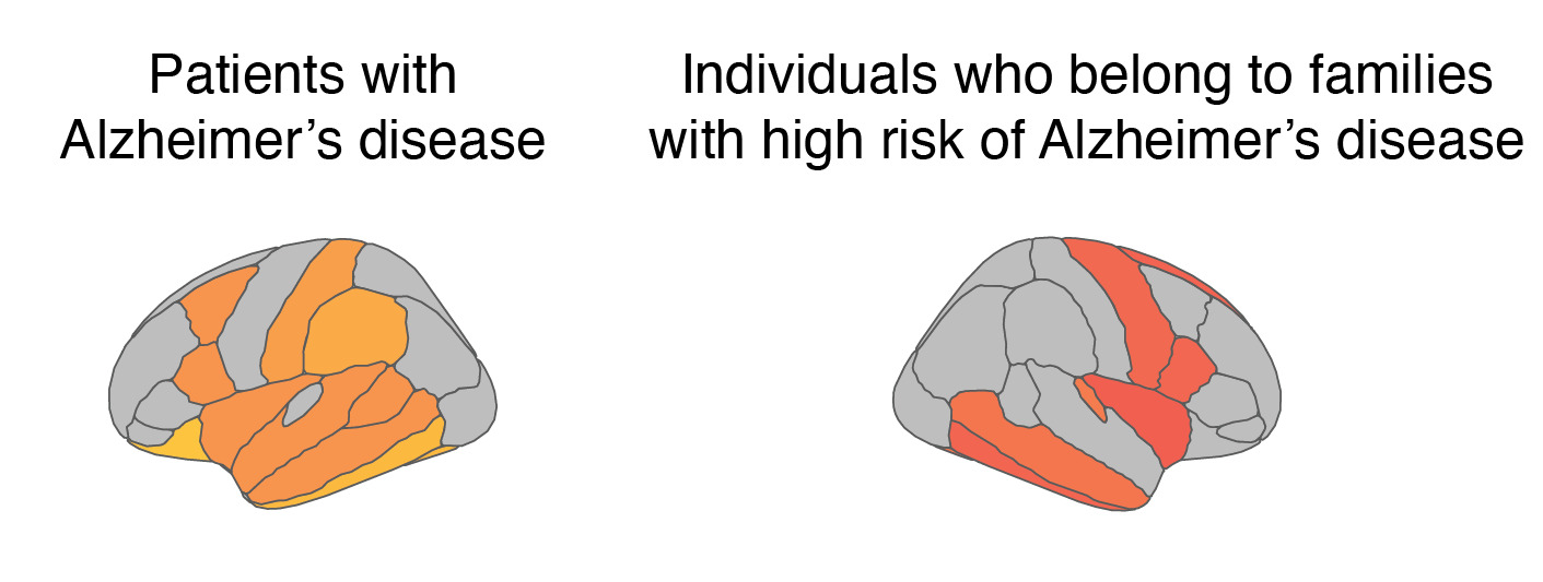
"This study included the participation of patients with mild cognitive impairment and Alzheimer's disease, as well as individuals belonging to families with a 50% risk of developing the hereditary form of Alzheimer's disease. All participants underwent comprehensive examinations using advanced brain imaging techniques to map the presence of astrocyte activation in the brain and blood tests. By comparing the cell activation depicted in the brain with blood test results for specific proteins, such as GFAP, we could evaluate if there was any connection between brain changes and the levels of the blood-based marker for astrocyte activation. By also analyzing the results from repeated examinations of the same individuals, we gained insight into the time course of activation of astrocytes."
Publication
”Tracking reactive astrogliosis in autosomal dominant and sporadic Alzheimer’s disease with multi-modal PET and plasma GFAP”, Konstantinos Chiotis, Charlotte Johansson, Elena Rodriguez-Vieitez, Nicholas J Ashton, Kaj Blennow, Henrik Zetterberg, Caroline Graff, Agneta Nordberg. Molecular Neurodegeneration, online September 12, 2023, doi: 10.1186/s13024-023-00647-y.
Funding
Swedish Research Council, Swedish Foundation for Strategic Research (SSF), Region Stockholm, Brain Foundation, Alzheimer's Foundation and The Center for Innovative Medicine (CIMED).