New exhibition about Lennart Nilsson on KI Campus Solna

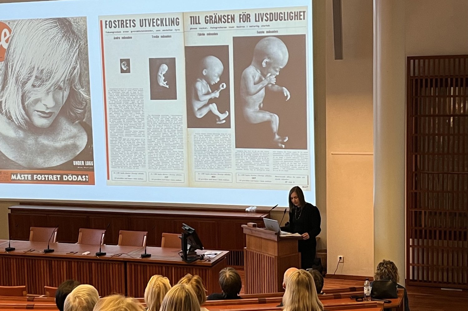
Photographer Lennart Nilsson became world famous for his images from the inside of the body. Foetuses during different stages of development and microscopically small cells could be photographed thanks to his electron microscope. Lennart Nilsson's pictures are now historic and many were taken when he worked at Karolinska Institutet.

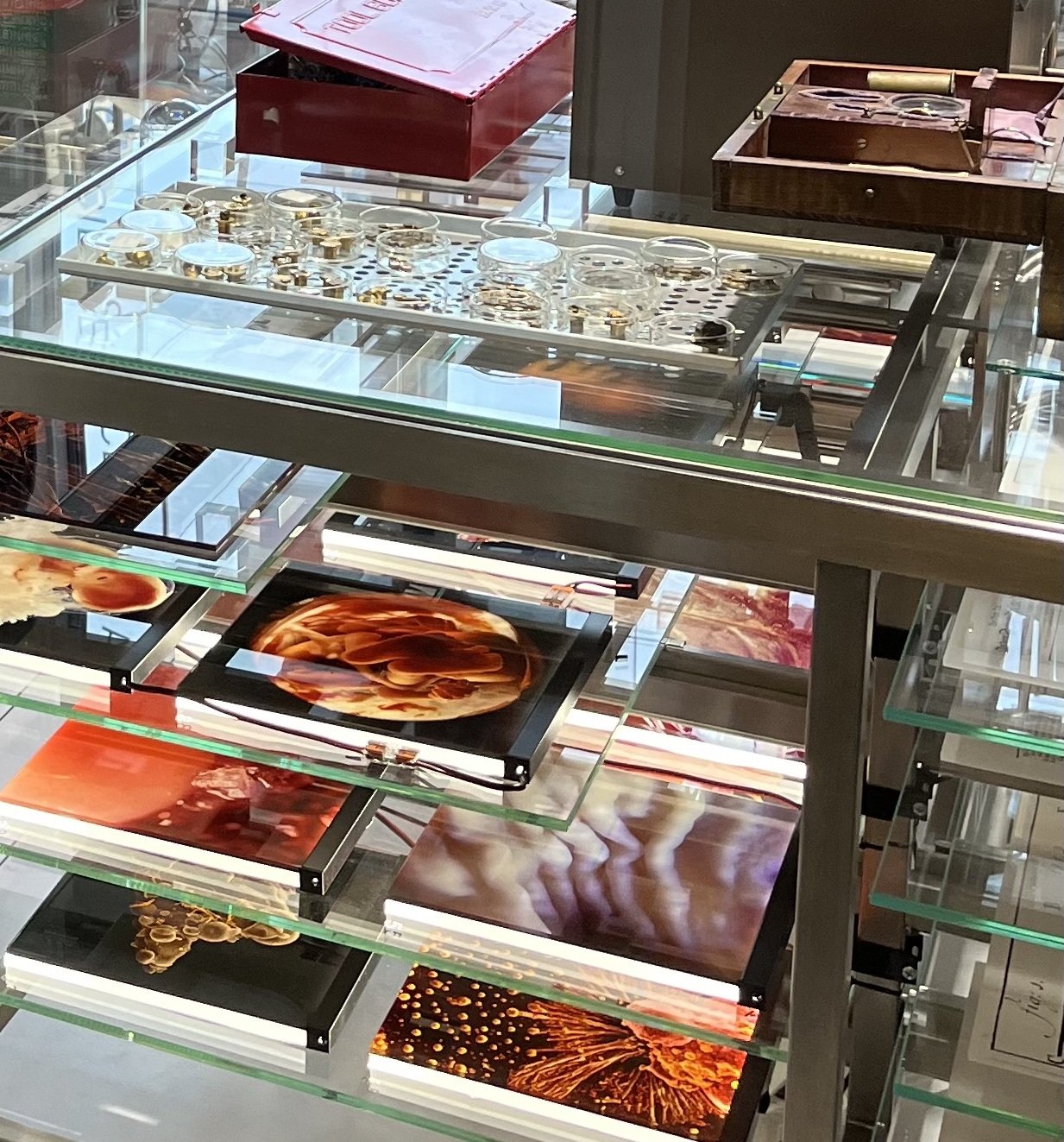
A new exhibition about Lennart Nilsson's works is now opening. The exhibition Art in Science – Science in Art. Lennart Nilsson and KI will be on display in the entrance to the Admin Building at Nobels väg 5 from Tuesday 14 May.
The exhibition uses material from Lennart Nilsson's last laboratory, which was taken care of by the History and Cultural Heritage of Medicine (Medicinens Historia och Kulturavr, MHK) in the spring of 2020. The initiator of the exhibition is Olof Ljungström, docent in the history of science and ideas, and head of unit at MHK.
“Lennart Nilsson and his work with electron microscopy and photography were groundbreaking, both technically and artistically. At the same time, his images are part of a much longer history in which science has used optics and light to be able to see and understand things that cannot be seen with the naked eye,” says Olof Ljungström.

The exhibition includes one of the electron microscopes Lennart Nilsson used, and a selection of the thousands of photographs he took. There is also an interactive screen where visitors can see into the human body for themselves.
The second part of the exhibition shows the history that Lennart Nilsson's pictures are linked to, with objects and images from the 1600s onwards, from the collections of KI and the Hagströmer Library.

Book a guided tour
The exhibition is open between 9 am and 3 pm on weekdays. Address: Nobels väg 5, KI Campus Solna.
To book a guided tour, please email olof.ljungstrom@ki.se
