New core facility offers world class imaging technique
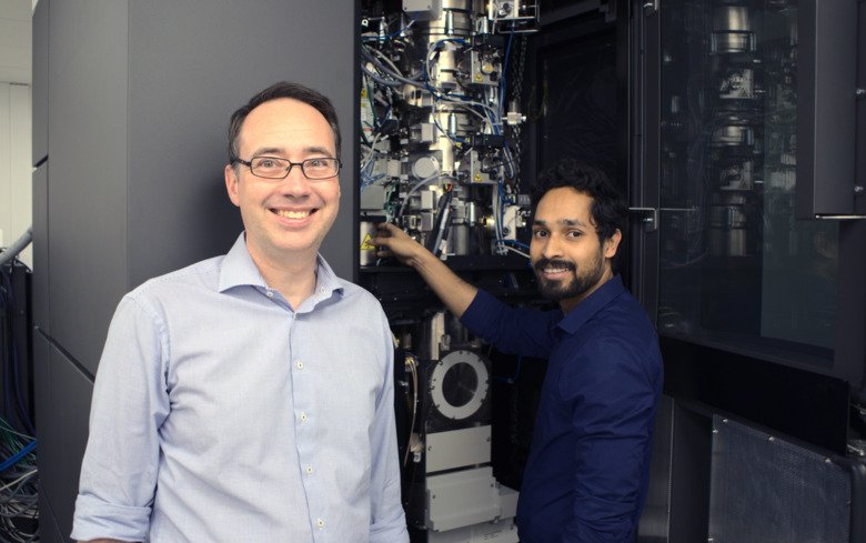
The corona pandemic put a stop to the planned inauguration-reception this autumn, but now Karolinska Institutet's new 3D-EM core facility is fully operational after all. In the basement of Wargentin House on Solna campus there are several cryogenic electron microscopes of the latest models, that can be used by both KI researchers and external customers. Martin Hällberg, PI at the Department of Cell and Molecular Biology and director of the 3D-EM facility, is enthusiastic.
What exactly have you installed?
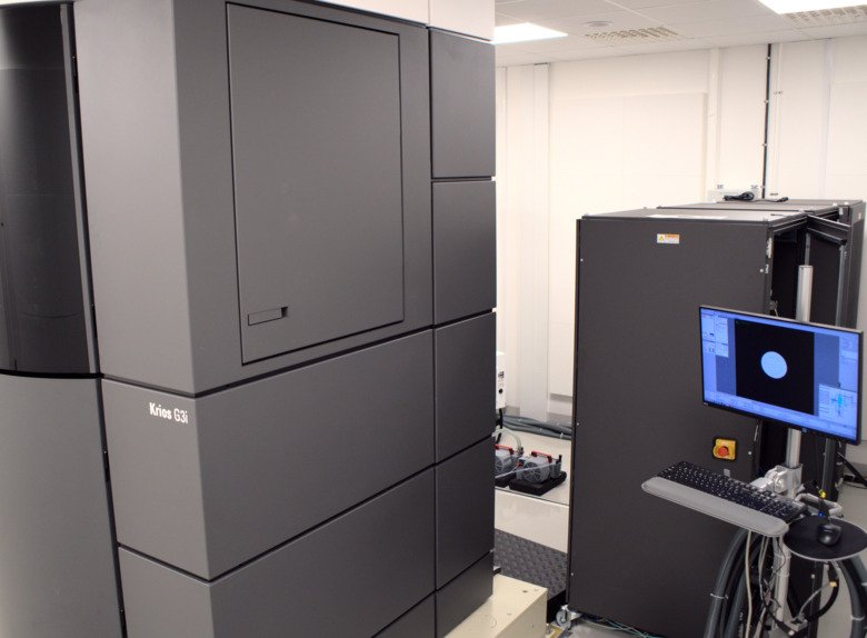
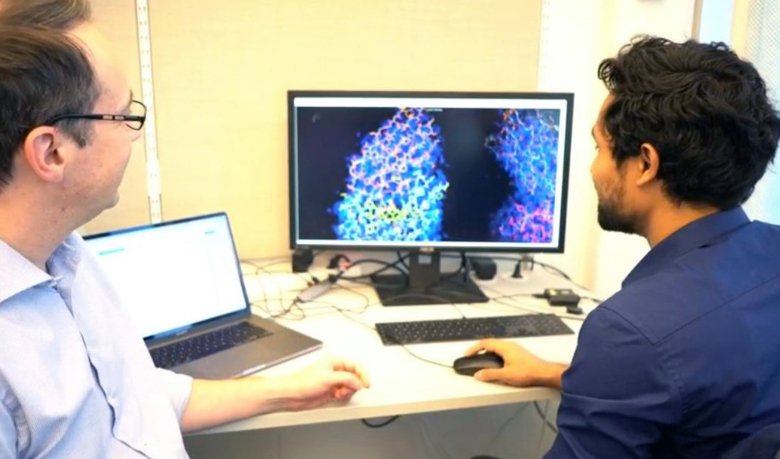
“We have several microscopes, but the top microscope, ‘Krios’, is the best on the market. It produces high resolution 3D images, which is enough for scientists to see, for example, the molecular structure of a protein complex in its natural state. Since the image is taken when the sample is instantly frozen, it can be seen in its native state, untainted by artificial treatments. This is equipment that makes it possible for scientists to conduct studies of a quality that’s accepted by the best journals.”
Has it already helped KI researchers become published?
“Yes, including a study on how spinal cord stem cells can be induced to form new neurons in mice with spinal injuries. Two studies on COVID-19 have also been published. In the first, researchers used data collected on the 3D-EM Krios to see how a tiny antibody attaches itself to a surface protein on SARS-CoV-2, blocking the ability of the virus to enter cells. This study was done on alpacas. The second study shows how a synthetic antibody called Sybody 23 can bind to a spike protein that helps the SARS-CcoV-2 virus to bind to cells.”
How much has it cost?
“The equipment budget adds up to around SEK 60 million and is financed with KI’s infrastructure funds. Most of the equipment has now been purchased and commissioned. It has taken years to get the facility up and running and we hit a few snags along the way. For instance, we only found out late in the building phase that the new metro line, which is being laid along Solnavägen, would involve blasting rather than drilling in this area. It took quite some time and effort to find a solution to the problem."
“COVID-19 also caused us problems as engineers couldn’t come here from the Netherlands and we had to severely cut back on user training. Now in the late autumn we’re also seeing a drop in the number of samples coming from the users, probably because COVID-19 has put a stop to much of the sample preparation. In the late spring and summer there were plenty of samples, many of which had been produced before the pandemic.”
How can researchers make use of this new facility?
“Researchers at KI pay a reduced user fee and receives training for free. The facility is also available for researchers from other universities and private companies, but they of course will have to pay more. Those interested can reach us via our website. They’ll then get training on one of our simpler microscopes to learn the technique.”
“Something we are very happy to be able to offer is excellent equipment for preparing samples, which can be frozen and made into successively thinner slices. All this we can help with. There are different levels of difficulty in the preparation of samples, the trickiest ones often requiring a multitude of trials before something useful can be obtained. We jokingly call these techniques ‘trial and horror’, but everything is moving forward and becoming more and more automated and predictable in terms of the quality you’re eventually going to attain.”
What researchers are your main customers?
“At first we had a lot of biochemists wanting to find out the molecular appearance of purified protein complexes in order to understand how they work. During the winter, we’ll be extending and completing the installation of sample preparation techniques that make it possible to look inside instantly frozen cells using cryogenic electron microscopy. It’s been only in the past few years that we’ve been able to study native cell material and tissue samples. Before this, you had to plastic-embed the material in the lab and stain it with heavy atoms, after which you can risk that natural structures of the cell either destroyed or corrupted. This technique is making rapid advances around the world, and now KI is on board too. Thanks to these developments, we’re going to see a cell-biology revolution in the coming years.”
Facts about cryo-electron microscopy
Three scientists, Jacques Dubochet, Joachim Frank and Richard Henderson, received the 2017 Nobel Prize in chemistry for their contributions to cryogenic electron microscopy. The method involves fixing the sample as it is submerged into liquid ethane; by imaging the sample from many different angles, scientists can then assemble a three-dimensional image, allowing them to look inside cells and understand their workings at a molecular level.