Neurons specialised in encoding sound emerge before birth
Distinct neuron types in the auditory organ are necessary for encoding different features of sound and relaying them to the brain. Researchers at Karolinska Institutet provide evidence of an early, neuronal activity-independent, emergence of the different subtypes of auditory neurons, prior to birth in mice. The findings have recently been published in Nature Communications.

Previous studies have provided ambiguous results on whether the different subtypes of auditory neurons emerge during prenatal or postnatal development, with in the latter case, a possible role of neuronal activity in generating their diversity. In this new study, researchers demonstrate that the fate of auditory neuron subtypes is under genetic control in the prenatal period, and reveal the complex molecular networks controlling their genesis.
Why are these results important?
“Our findings demonstrate that most aspects directly linked to the functional diversity of auditory neurons seem to be intrinsically, molecularly defined before birth,” says Francois Lallemend, Principal researcher at the Department of Neuroscience, and last author of the article. “This has implications on how we can interpret peripheral auditory (dys-)functions at birth but also for devising ways to convert neuron types in pathological conditions or congenitally deaf cochlea, and hopefully improve hearing.”

How did you perform the study?
“We mapped the gene expression of auditory neurons over several days of development, using single-cell transcriptomics approach, and used advanced analytical tools to reveal a hierarchical tree of neuronal diversification,” says Saida Hadjab, principal researcher at the Department of Neuroscience and co-last author of the article. “We could explore in detail, at single cell level, the dynamic of molecular developmental changes as neuron types were diverging and their identities were progressively acquired.”
“We publish the whole molecular program that defines the diversification events. We then use mouse genetics to functionally evaluate this program, and eventually compare it with published datasets of deafness genes, revealing deafness genes associated with neuronal differentiation”, she continues.
The next step
Several years ago, the researchers showed that neuronal diversity in the peripheral auditory system had a strong molecular basis, providing ways to manipulate each subtype to better understand their role in hearing.
With this study they demonstrate the molecular programmes that set this diversity before the peripheral auditory system is even active. “Our next step is to explore if and how this diversity is affected in pathological conditions, for instance after noise trauma or aging, and use our new molecular knowledge and tools for resetting auditory neuron function and recovering normal hearing”, says Francois Lallemend.
The study was financed by the Swedish Research Council, Knut and Alice Wallenberg Foundation, Karolinska Institutet, the Ming Wai Lau Foundation, the Brain Foundation and Tysta Skolan foundation.
Publication
Single-cell RNA-sequencing analysis of the developing mouse inner ear identifies molecular logic of auditory neuron diversification.
Petitpré C, Faure L, Uhl P, Fontanet P, Filova I, Pavlinkova G, Adameyko I, Hadjab S, Lallemend F
Nat Commun 2022 Jul;13(1):3878
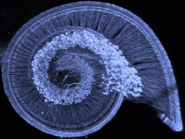
The rotations show the shape of the structure of the hair cells and spiral ganglion cells from more perspectives. Then, zoom to the hair cells / spiral ganglion processes / spiral ganglion neurons at the basis of the cochlea and, as on a ladder, the picture slowly moves upper and upper towards the apex of the cochlea. This spatial organization of hair cells in the Corti-organ is important in the discrimination of lower and higher frequency voices. Then, more zoom to the apical inner hair cells.