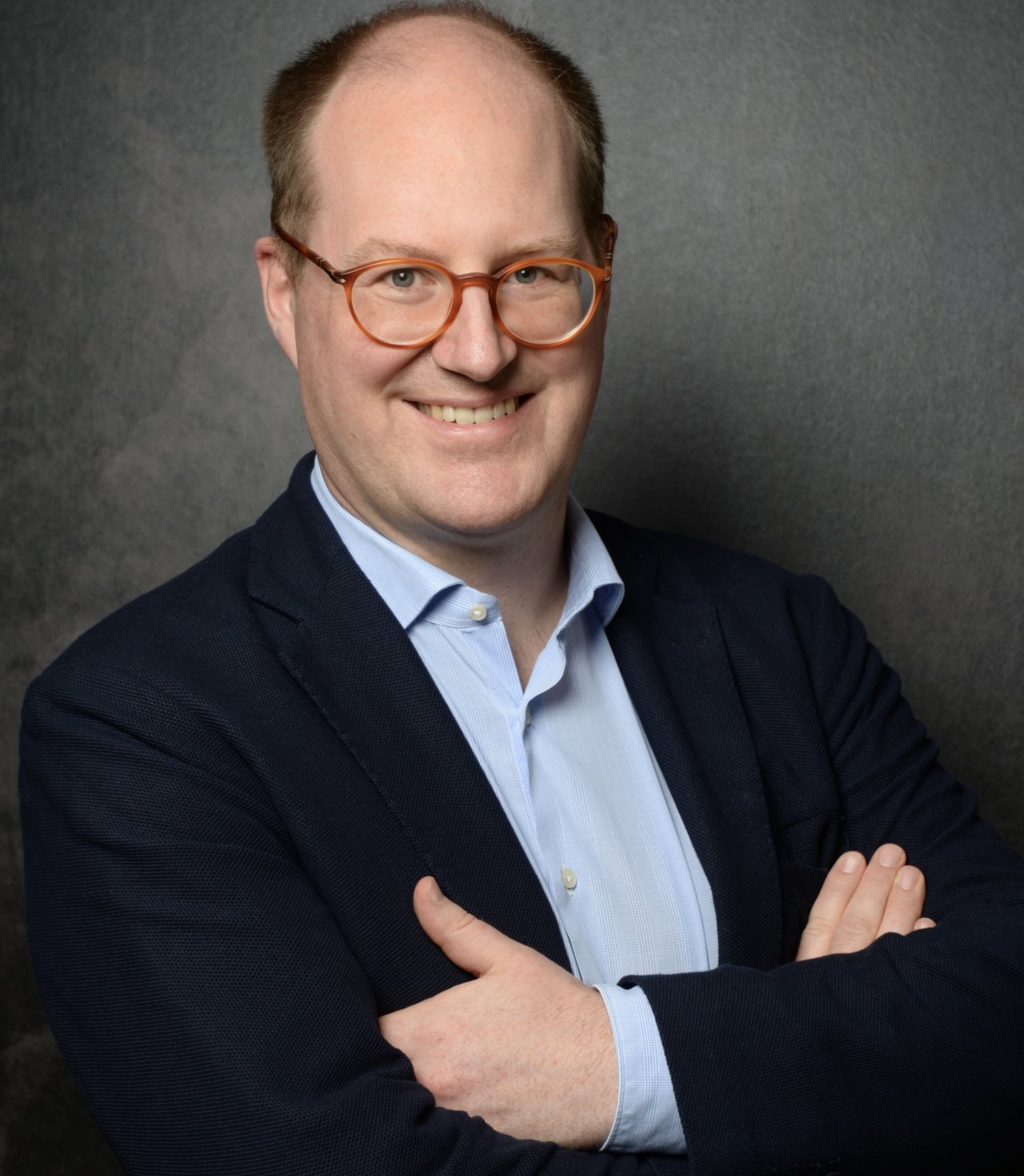
Maximilian Ackermann receives the 2025 Lennart Nilsson Award
Dr. Maximilian Ackermann, currently working as a Professor of Pathology at the RWTH University Clinics Aachen and Helios University Hospital Wuppertal, Germany, and as anatomist at the Johannes Gutenberg University Mainz is awarded the 2025 Lennart Nilsson Award for his outstanding contributions to scientific imaging, revealing critical insights into diseases, such as COVID-19 and cancer.
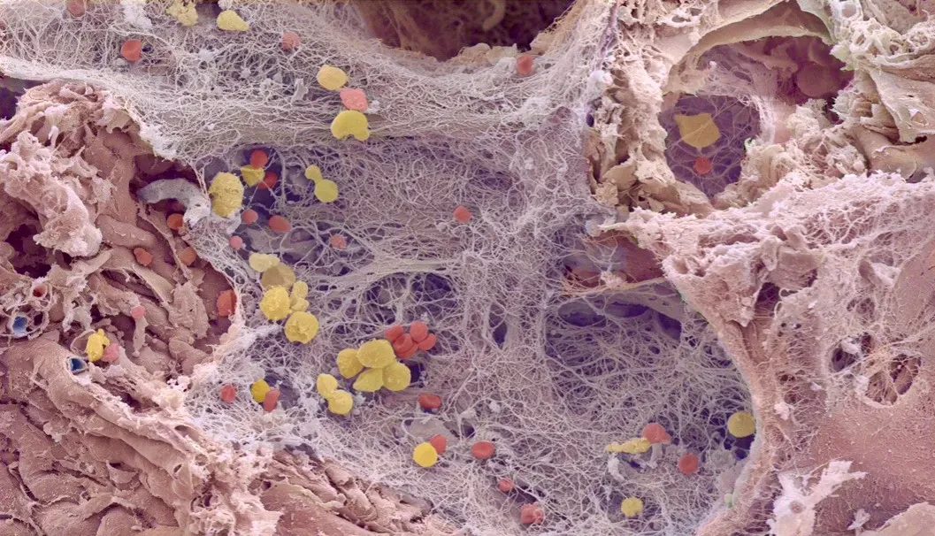
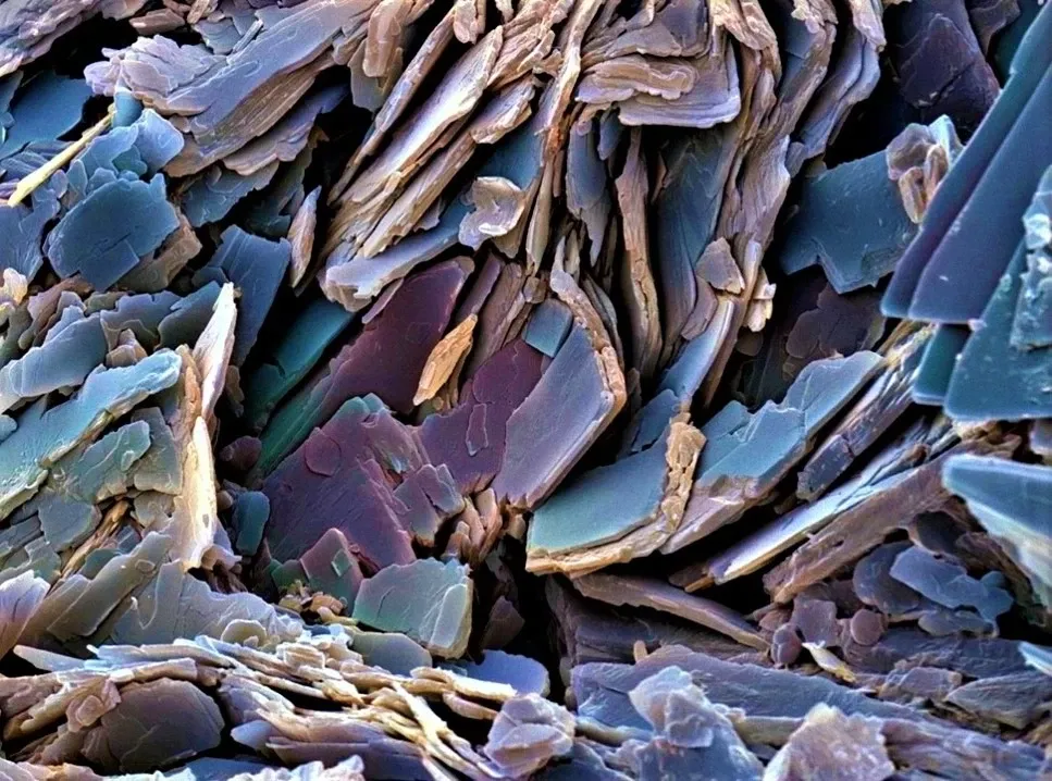
Dr. Ackermann specialises in pathology and anatomic pathology and has a passion for artistic photography. His images provide a glimpse into both the beauty of human anatomy and the darker aspects affected by disease, while also illustrating new scientific discoveries related to the condition.
“The morphology of two-dimensional tissue sections is crucial for clinical pathologists and anatomists to make far-reaching diagnoses for our patients, such as in the case of cancer,” says Dr. Ackermann. “In doing so, I am always interested in how pathological changes in cancer or inflammation affect tissue in three dimensions. The principle of ´form follows function´, a fundamental tenet in modern architecture, similarly applies to the anatomy of tissue.”
Bridging radiology and pathology
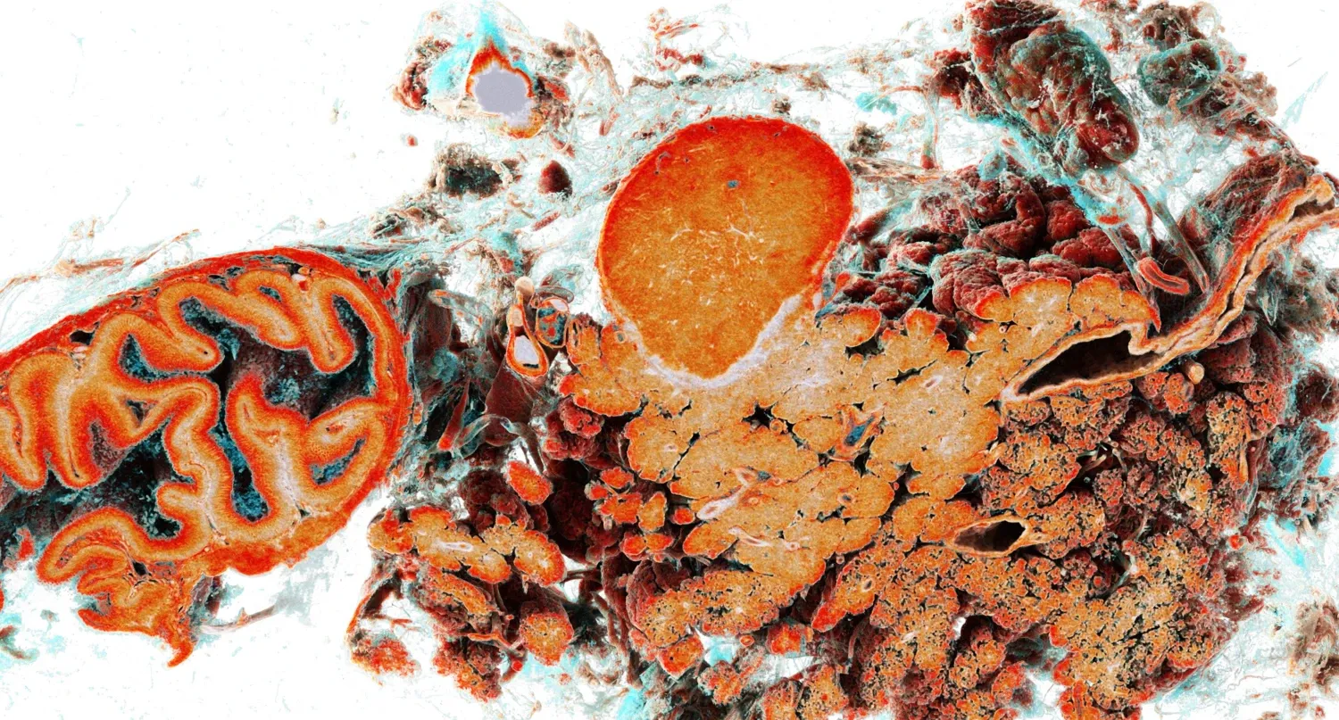
Ackermann’s work contributes to advancing our understanding of complex biological structures and disease. It includes developing and applying advanced techniques like hierarchical Phase-Contrast Tomography (HiP-CT), which is currently being integrated into clinical diagnostic processes.
“This means that we are generating results with our classic microscope and supplementing them with the three-dimensional data from HiP-CT. For example, in our HIMALAYA project, we analyse prostate carcinomas holistically. The same is true for breast carcinomas post chemotherapy. HiP-CT is an excellent bridging technology that brings together specialities such as radiology and pathology. It will undoubtedly provide substantial support in the characterisation of diseases and in AI-based image recognition in radiology.”
Extraordinary scientific photography
The Lennart Nilsson Award Foundation was established in 1998 in recognition of the world-renowned Swedish photographer Lennart Nilsson and his extraordinary body of work. The annual award recognises outstanding contributions within the realm of scientific photography.
“It is an extraordinary honour to receive this distinguished award, which melds art and science in a unique way and boasts a long tradition of exceptional scientists who have pushed the boundaries of their respective disciplines,” he says.

Dr. Ackermann's award-winning work was conducted at the European Synchrotron Radiation Facility (ESRF) in Grenoble, France.
The award ceremony will be held in conjunction with the installation ceremony for new professors and laureates in Aula Medica at Karolinska Institutet, Solna, Sweden on October 9th, 2025.
The jury’s citation
“The Lennart Nilsson Award 2025 is presented to Dr. Maximilian Ackermann. Dr. Ackermann is honoured for his outstanding contributions to scientific imaging, embodying the spirit of Lennart Nilsson by making the invisible visible. His pioneering work, particularly in developing and applying advanced techniques like hierarchical Phase-Contrast Tomography (HiP-CT) and artistic scanning electron microscopy, contributes to advancing our understanding of complex biological structures and disease.
Dr. Ackermann's visually stunning and scientifically significant images have revealed critical insights into diseases such as COVID-19 and cancer, presenting scientific breakthroughs to the world in beautiful, unique, and powerful ways. He has an extraordinary ability to translate intricate scientific concepts into compelling visual narratives.”