Science becomes art

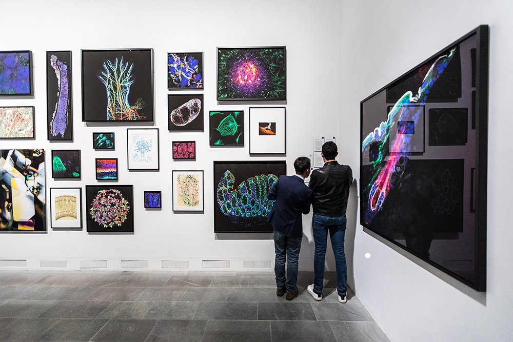
On the 31 October the exhibition The Invisible Body opened at Sven-Harry’s Art Museum in Stockholm. A number of researchers at KI are represented by pictures from their own research.
The new art exhibition The Invisible Body (Den osynliga kroppen) narrates the story of ongoing medical research from the perspective of medical images.
“In my role as a communicator, I have often experienced the difficulty in finding a way to talk about research in a comprehensible manner yet without oversimplification. The idea for this exhibition came when a researcher showed me a beautiful image obtained during her work. We soon realised that, through her picture, we had discovered an excellent way of discussing her research. Here, beautiful images become an entirely new point of entry to science,” explained Mona Norman, project manager at the Ragnar Söderberg Foundation, at the exhibitions opening.
The image was taken in Maria Kasper’s research group at KI’s Department of Biosciences and Nutrition and shows an area of the skin that is particularly susceptible to cancer. Her laboratory studies follicular stem cells in order to understand their link to wound healing and cancer.
“The opportunity to gain access to an advanced microscope that makes it possible to take these types of pictures and to among other things study the development of individual cancer cells, was the reason why I chose to locate my research at KI,” says Maria Kasper who attended the vernissage together with doctoral candidate Karl Annusver, the photographer behind the image.
The exhibition collects a number of works that depict subjects invisible to the naked eye, giving us new insights into how the healthy and diseased body functions. However, these images are also things of beauty. They are artworks that, after the exhibition closes, will be auctioned to raise funds to support research. And, according to Saida Hadjab at KI’s Department of Neuroscience, there are similarities between art and science.
“Creativity is a vital ingredient in the work of both artists and researchers.”
Saida herself has two pictures in the exhibition and is one of the project’s two scientific coordinators. Her own great interest in photography has proved an important motivational factor behind her research work at the microscope.
“I search for images that are both elucidatory and beautiful. A beautiful picture has a greater ability to convey its message,” she says.
The exhibition runs until 7 January 2018 and is an initiative from Ragnar Söderberg Foundation in collaboration with Sven-Harry’s Art Museum. It includes pictures from Hagströmer Medico-Historical Library, Lennart Nilsson Photography and researchers at a number of universities. In conjunction with the exhibition, a number of popular science lectures will be held. Learn more about the exhibition here.
In conjunction with the exhibition, a number of popular science lectures will be held. Learn more about the exhibition.
The full list of researchers from Karolinska Institutet that contributed to the exhibition
- Eduardo Guimaraes // Christian Göritz lab
- John Schell // Fredrik Lanner lab
- Carmelo Bellardita // Ole Kiehn lab
- Tomas McKenna // Maria Eriksson lab
- Elisa Floriddia // Fanie Barnabé-Heider lab
- Javier Calvo Garrido // Anna Wredenberg lab
- Anders Hånell // Histology course, Neuroscience department
- Cajsa Classon // Liv Eidsmo lab
- Saida Hadjab // François Lallemend lab
- Karl Annusver // Maria Kasper lab
- Jan Krivanek // Igor Adameyko lab
- Shigeaki Kanatani & Laura Heikkinen // Per Uhlén lab
- Katarzyna Malenczyk // Tibor Harkany lab
- Sten Linnarsson // Sten Linnarsson Lab
- Matthijs C. Dorst // Gilad Silberberg lab
- Ada Delaney // Camilla Svensson lab
- Margherita Zamboni // Jonas Frisén lab
- Laura Comley // Eva Hedlund lab
- Nicola Crosetto // Nicola Crosetto lab
- Sebastian Hildebrand // Fredrik Lanner lab
- Daniel Fürth // Konstantinos Meletis lab
- Sofie Ährlund-Richter // Marie Carlén lab
- Tomas McKenna // Maria Eriksson lab
- Benjamin Götte // Gerald McInerney lab
- Anna Rising Lab
