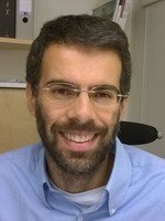
Grant from the Swedish Foundation for Strategic Research to Nicola Crosetto
Congratulations Assistant professor Nicola Crosetto, whose project” Integrated VISualization of Intra-Tumor Heterogeneity” was funded with 33 million SEK by the Swedish Foundation for Strategic Research!

What's the purpose of the project?
- The project has two main goals; to generate 3D tumor maps that integrate classical histopathology, gene expression and mutation data from multiple tissue sections of solid tumors and to apply a set of tools from a recently developed field of mathematics named Topological Data Analysis (TDA), with the goal of integrating high-dimension genomic, transcriptomic and imaging data obtained from tumor specimens, and represent this complex information in a condensed, synthetic form that can be easily understood by pathologists and clinicians.
These tools can be applied both to the data that we will be generating in aim 1, but also to already available cancer data, for example data from the international Cancer Genome Atlas (TCGA) consortium.
Why is that important?
- We are in a time when more and more so-called precision drugs are available in the clinic to target specific alterations of cancer cells, but at the same time many patients still develop resistance to new drugs at some point during therapy. Targeted therapies often give spectacular results at the onset of therapy, but then invariably fail due to the onset of resistance. Although in many cases we still don't know the exact molecular mechanisms of resistance, we know that there is a key feature of cancers that makes them prone to "learn" how to resist our attempts to kill them: this is so-called tumor heterogeneity, or in other words the fact that a tumor is typically composed of different tumor cell types mixed to normal cells like lymphocytes and fibroblasts, which continuously compete or cooperate with each other and evolve under the selective pressure of therapy. Pretty much like species evolve in Darwinian evolution under the selective pressure of the environment. In this process, sooner or later a subclone of cells that learnt how to resist treatment will emerge, causing the treatment failure that we see in the clinic.
It is therefore essential that we get to know the principles and rules that govern this heterogeneity and evolutionary dynamics inside tumors. Unfortunately, it is not possible at present to do this in real time in a patient, however what we can do is trying to reconstruct this dynamics by looking at many different parameters in many different regions of tumor samples by creating rich tumor maps, pretty much like ecologists create maps of where the different species and resources are located in the ecosystem they study. So, we need to start a new field of research which I propose to call Tumor Topography, and our project goes precisely in this direction.
How could your project contribute to the improvement of people’s health?
- There are at least two ways by which I see that our project will benefit cancer patients and more broadly society in the next five years. The first is by improving tumor classification and helping pathologists and clinicians to precisely classify each individual tumor, so that the most appropriate drug or drug combination can be offered to the patient. As soon as we demonstrate that our quantitative approach is able to reliably classify different types of cancer and capture meaningful information that would be otherwise "invisible" to the human eye – for instance prognostic patterns hidden in large omic datasets or quantitative prognostic features embedded in histopathological images that cannot be detected by eye – we can start translating it in the clinic. My dream is to see, in five years from now, that quantitative, computer-aided tumor topography is implemented as part of routine cancer diagnostics at Karolinska University Hospital and ideally in other hospitals across Sweden.
The second way by which I believe our project will positively impact society in the long term is by providing for the first time a thorough understanding of tumor architecture in 3D at high resolution. So far, we can use powerful techniques such as computer tomography to get a 3D picture of diseased tissues and organs, but we cannot see for example how different cell types and blood vessels are spatially organised in the whole tumor volume. This is extremely important, for example to model how drugs can reach various parts of a tumor, or to identify small populations of drug-resistant cells in a small region of the tumor that would go likely undetected in conventional 2D histopathology. I think that being able to image the morphology and molecular composition of tumors in 3D at single-cell resolution will truly revolutionise the way we model this complex disease, and give us new inspirations for how to attack it more effectively.
In your opinion, how important is “big data” research for medical research?
- I would say it is essential. In the past decade, the IT revolution together with the emergence of omic technologies such as next-generation sequencing have literally created an explosion of digital medical information, including digital clinical records, digital pathology, clinical genomics, and computer-assisted imaging. This massive flow of information will grow at an even faster pace in the coming years, as more and more high-throughput and information-rich technologies become part of routine diagnostics. Therefore, we are facing an urgent need to develop quantitative approaches that can integrate all these "big data" and synthesise them for clinicians in a form that can help them to make the most appropriate decisions in the interest of their patients.
I believe we also need to radically reform the way we train the next-generation of physicians and healthcare providers, for example by introducing in their curriculum obligatory courses in computer programming and a stronger training in statistics and data science, but also by creating inter-disciplinary programs where clinicians are trained alongside molecular biologists, computer scientists, and technologists.
