Cellular architecture of lesions in MS now mapped out
Using advanced methodology, scientists in Sweden were able to reveal at the cellular level how lesions in multiple sclerosis develop. The new results are presented in the journal Cell by researchers from Karolinska Institutet and Stockholm University.
Over 1,8 million people worldwide are diagnosed with multiple sclerosis, MS. In this disease, the body's immune cells attack the cells that form myelin, the so-called oligodendrocytes, which belong to the group of glial cells. Without myelin, signals between nerve cells cannot travel as fast as usual, resulting in symptoms such as reduced sensation and lack of coordination. MS is characterized by lesions in the brain and the spinal cord.

"We wanted to understand which cells are part of the lesions and their dynamics over time," says Petra Kukanja, co-first author in the study and PhD student at the research group of Professor Gonçalo Castelo-Branco, at Department of Medical Biochemistry and Biophysics, Karolinska Institutet.
The researchers used a technology called in situ sequencing, developed in the research group of Professor Mats Nilsson, at Stockholm University. This involves analyzing and identifying cells that are part of a tissue section by reading which genes are active in a particular cell. This pattern reveals both how different cell types are arranged in the tissue, in this case the spinal cord, and how the cells interact with each other. To study how the lesions develop, samples were taken at different times from mice experimentally induced with MS-like-symptoms and from human MS patients.

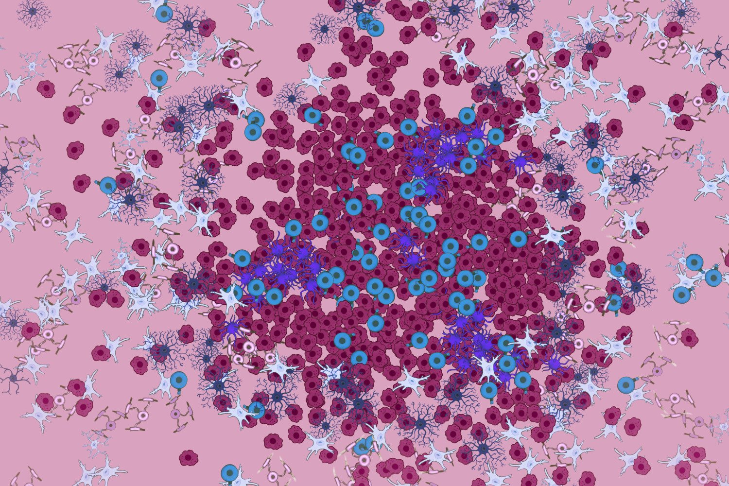
"We analyzed simultaneously 239 genes and saw that active lesions in mouse were built up centrifugally in two dimensions, with immune cells in the middle and different types of glial cells around them," says Christoffer Mattsson Langseth, also co-first author of the study, and PhD student at the Department of Biochemistry and Biophysics, Stockholm University at Professor Mats Nilsson’s group.
In the mice, it was possible to see that the lesions first appeared in the spinal cord and then spread towards the brain. In samples of the spinal cord from four deceased MS patients, 260 genes were simultaneously analyzed and the cellular architecture of the lesions could be determined. The authors also found new lesions and new sub-structures within the lesions.
Victims of immune cell attacks
Previously, oligodendrocytes have mainly been seen as victims of immune cell attacks.
“The fact that they are active in the outer regions of lesions but also in the entire brain and spinal cord raises the question of whether they dampen the disease or drive it,” says Petra Kukanja.
In the next step, the researchers want to use the same methodology to analyze samples from more MS patients, as the disease is very heterogeneous. Another question is what the lesions look like when patients have received different types of treatments.
The two researchers highlight the extensive and fruitful collaboration between their research groups, which has been ongoing since 2019 and to which they have contributed their respective expertise - from Karolinska Institutet on the cell biology behind MS and from Stockholm University with their pioneering technology in situ sequencing and new methodology for complex image analysis.
The study was initially funded by The Strategic Research Area Neuroscience (StratNeuro) and throughout the project by the Swedish Brain Foundation, the Swedish Cancer Society, the Göran Gustafsson Foundation for Research in Natural Sciences and Medicine, the Swedish Society for Medical Research, Olav Thon Foundation, Ming Wai Lau Centre for Reparative Medicine, the Swedish Research Council, the Chan Zuckerberg Initiative, the Erling-Persson Foundation and the Knut and Alice Wallenberg Foundation, European Union (European Research Council). Markus M. Hilscher and Mats Nilsson owned shares in CartaNA AB, which is part of 10x Genomics. Mats Nilsson is a former scientific advisor for 10xGenomics.
Publication
”Cellular architecture of evolving neuroinflammatory lesions and multiple sclerosis pathology”, Petra Kukanja*, Christoffer M. Langseth*, Leslie A. Rubio Rodríguez-Kirby, Eneritz Agirre, Chao Zheng, Amitha Raman, Chika Yokota, Christophe Avenel, Katarina Tiklová, André O. Guerreiro-Cacais, Tomas Olsson, Markus M. Hilscher, Mats Nilsson, Gonçalo Castelo-Branco, Cell, online March 20, 2024, Cell, doi: 10.1016/j.cell.2024.02.030